MEDICALHOLODECK®. Surgery, Anatomy, Medicine, Education, and DICOM Imaging in VR. For Medical Students, Nurses, Doctors and Surgeons. - nooon34
See how Medicalholodeck supports neurosurgery in civil war, how spatial imaging aids spine surgery, and how iPads enhance radiology workflows.

View the Presentations


MEDICALHOLODECK®. Surgery, Anatomy, Medicine, Education, and DICOM Imaging in VR. For Medical Students, Nurses, Doctors and Surgeons. - nooon34
RecordXR is the spatial editor for XR content production. This powerful tool turns any XR session into fully interactive, immersive content that can be recorded, edited, and shared across devices—without the need for coding or video production skills.


Whether you're a creator, educator, or trainer, RecordXR Studio enables you to build your own immersive content library, capturing every movement, interaction, and conversation in 3D space. Welcome to a new era of content creation—designed natively for extended reality.

What Is RecordXR Studio?
RecordXR Studio is a 4D recording and editing tool built into the Medicalholodeck platform. It captures everything that happens inside an XR session—including object manipulation, user motion, tool usage, audio, and environment navigation.


But unlike traditional screen recording, it doesn’t just record what you see—it records what you do. Once captured, your session becomes a fully replayable spatial experience. You can review it from any angle, at any time, in real 3D.

Why It’s a Game-Changer for XR Creators
Spatial computing is here. But up until now, creating shareable XR content has required specialized software, manual video capture, or custom development. RecordXR Studio changes that.

With a single click, you can:
  • Capture immersive learning modules
  • Document interactive workflows
  • Record virtual walkthroughs or training demos
  • Save team discussions and collaborative sessions
  • Build modular 4D lessons or experiences

The best part? All of this can be replayed, edited, and shared—in full spatial format—across a wide range of devices.
MEDICALHOLODECK®. Surgery, Anatomy, Medicine, Education, and DICOM Imaging in VR. For Medical Students, Nurses, Doctors and Surgeons. - nooon34
March 15, 2025 | 14:00 – 16:00 CET / 09:00 – 11:00 EDT

More information

Register here for the free online conference

MEDICALHOLODECK®. Surgery, Anatomy, Medicine, Education, and DICOM Imaging in VR. For Medical Students, Nurses, Doctors and Surgeons. - nooon34
Medicalholodeck’s latest update to version 1.3.1 delivers new AI-powered segmentations, optimized volumetric rendering for very big datasets with high performance, and language support for Brazilian Portuguese.

New AI-Powered Segmentations

Medicalholodeck now additionally supports AI segmentation for Liver Vessels and Oculomotor Muscles, expanding its capabilities for medical professionals working in radiology, surgery, and medical education.

These new models, integrated from TotalSegmentator, allow users to efficiently analyze complex anatomical structures in PC VR and Remote Rendering environments.

With these additions, Medicalholodeck continues to provide a powerful tool for visualizing and interacting with medical imaging data in real-time.

In addition to the newly introduced segmentation models, Medicalholodeck already supports the following range of AI segmentation models, including:

⚡Main Structures⚡
⚡MRI Main Structures⚡
⚡Lung Vessels⚡
⚡Body⚡
⚡Head Glands & Cavities⚡
⚡Head Muscles⚡
⚡Neck Bones & Vessels⚡
⚡Neck Muscles⚡
⚡Coronary Arteries⚡
⚡Cerebral Bleed⚡
⚡Pleural & Pericardial Effusion⚡
⚡Hip Implant⚡
⚡NEW: Liver Vessels⚡
⚡NEW: Oculomotor Muscles⚡


The segmentation process is efficient and straightforward: import your DICOM data, select the appropriate AI segmentation model, and let the system process the structures automatically. Segmentation is fast, taking only 20–50 seconds for a standard-sized DICOM dataset.

Once the initial segmentation is complete, you can refine it by selecting or deselecting specific structures to focus on the critical details. This automation significantly reduces manual effort, enhances accuracy, and allows for a more precise and immersive study of anatomical relationships.

These advanced segmentation options are particularly valuable for hepatic surgeons, radiologists, ophthalmologists, and medical educators. Liver vessel segmentation supports preoperative planning and research, while detailed segmentation of the oculomotor muscles provides critical insights into orbital anatomy, essential for ophthalmic surgery and neuro-ophthalmology.

Medicalholodeck’s AI segmentation models already include Coronary Arteries, Cerebral Bleed, and Lung Vessels, offering a comprehensive toolset for anatomical analysis and surgical planning.

Higher Performance for Large Datasets
Medicalholodeck’s volumetric rendering has been significantly optimized, achieving a 20–40% increase in framerate on PC VR and Remote Rendering. This enhancement enables medical professionals to work with larger DICOM datasets more efficiently, ensuring smoother interactions and clearer, more detailed visualization of complex anatomical structures.

For radiologists, surgeons, and medical educators, this upgrade translates into reduced lag, improved image clarity, and a more immersive VR experience which is critical for accurate assessments and procedural planning. Higher frame rates allow for more intuitive interaction with patient data, whether analyzing volumetric scans, preparing for surgery, or teaching anatomy in 3D.

For more information, contact info@medicalholodeck.com
MEDICALHOLODECK®. Surgery, Anatomy, Medicine, Education, and DICOM Imaging in VR. For Medical Students, Nurses, Doctors and Surgeons. - nooon34
Medicalholodeck is now available on iOS, bringing its full suite of medical augmented reality applications to iPads and iPhones. With this launch, healthcare professionals, educators, and students can access Medicalholodeck’s medical visualization tools directly on their mobile devices. Additionally, it enables collaboration between users on VR headsets and iOS devices.


Advanced Medical Visualization on Mobile Devices
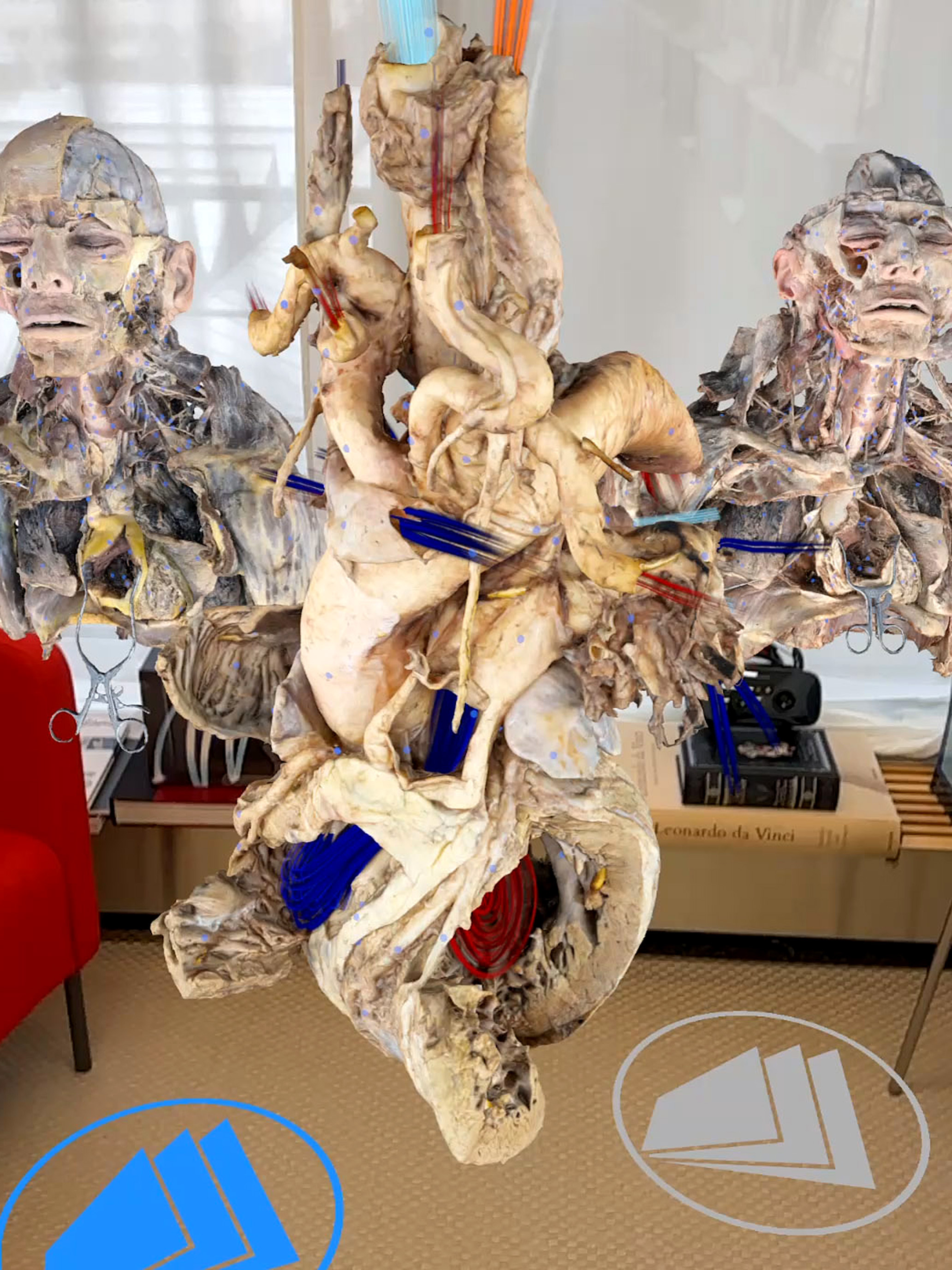
The Medicalholodeck suite offers a complete set of AR-powered applications designed for in-depth exploration, analysis, and interaction with medical imaging. Users can load patient data in DICOM format and view it in augmented reality, enhancing diagnostic and educational experiences. The platform also includes the Dissection Master XR and Anatomy Master 2 datasets, featuring high-resolution anatomical models for learning and teaching. With the addition of an iOS version, Medicalholodeck is now available not only on VR headsets but also on iOS mobile devices, expanding its use in hospitals and classrooms.

Expanding Beyond VR to Mobile Devices
Medical students can study detailed anatomical models and medical imaging on their own devices, improving their understanding of human anatomy and pathology. They can examine virtual cadavers, manipulate 3D structures, and compare healthy versus pathological conditions, enhancing their diagnostic skills. The ability to collaborate with peers and instructors using a combination of VR headsets and mobile devices further enhances the learning experience.


Healthcare professionals can review DICOM scans and 3D models remotely, facilitating better communication with colleagues and patients. This enables quick and effective discussions about surgical procedures, treatment planning, and case reviews without requiring access to traditional workstations. With support for mixed-device collaboration, professionals using VR headsets can now interact seamlessly with colleagues using tablets and smartphones.

Educators can integrate interactive AR tools into their teaching, making medical concepts more accessible and engaging. By presenting medical imaging and anatomical structures in AR, instructors can create a more immersive and interactive learning experience for students. Multi-device compatibility allows instructors to conduct virtual dissections and interactive demonstrations with participants joining from different platforms.

Clinicians handling patient information can use Medicalholodeck to review patient-specific DICOM scans directly from their mobile devices, ensuring they have access to crucial medical data at any time. The ability to interact with specialists using VR headsets enhances remote consultations and decision-making.


Use Cases in Detail
Surgical Planning and Patient Communication: Surgeons can use Medicalholodeck to review and explain complex cases to patients, offering a visual and interactive way to discuss procedures. The ability to display 3D reconstructions of patient scans helps improve patient understanding and trust in medical decisions. The mixed-reality capability allows surgeons to consult with colleagues using both VR headsets and mobile devices in real time.*

Radiology and Imaging Analysis: Radiologists can analyze and interpret DICOM images directly from their iOS devices, enabling remote consultations and second opinions. This is especially beneficial for specialists working across multiple facilities or in telemedicine scenarios. By integrating VR headsets into the workflow, radiologists can interact with 3D scans more intuitively while sharing insights with colleagues using mobile devices.

Medical Training and Education: With AR-enhanced anatomical models and medical imaging, students can engage in self-guided learning or participate in instructor-led virtual dissections. This approach helps bridge the gap between theoretical knowledge and practical application. The ability to work collaboratively across different devices ensures more inclusive and engaging educational experiences.

Download Now on iOS
With Medicalholodeck now available on iOS, users can experience a new level of accessibility and flexibility in medical AR. Whether for education, professional use, or research, this powerful tool brings medical visualization to the palm of your hand while supporting real-time collaboration between VR and mobile users.

Download Medicalholodeck on the App Store and explore the future of medical AR today.
MEDICALHOLODECK®. Surgery, Anatomy, Medicine, Education, and DICOM Imaging in VR. For Medical Students, Nurses, Doctors and Surgeons. - nooon34
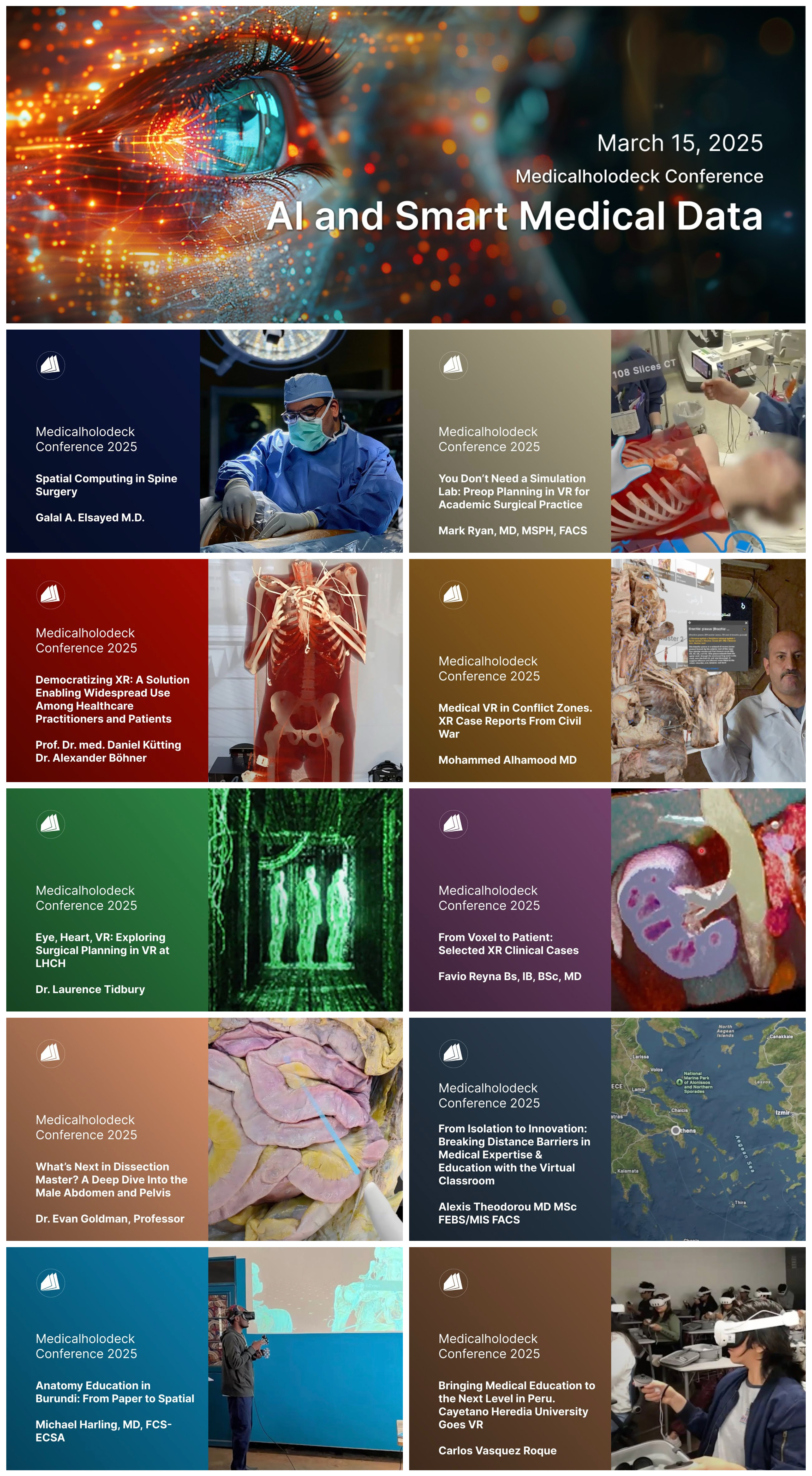
Medical AI and spatial technologies are reshaping surgery, clinical practice, and medical education. Join us on March 15, 2025, remotely or in Zurich, Switzerland for the Medicalholodeck Conference & Investors Day, where industry experts, surgeons, and investors will discuss and explore the future of AI-driven healthcare solutions.

The event is divided into two parts:

Part 1: The Medicalholodeck Conference will highlight the latest advancements in AI-powered surgical planning and smart medical data, featuring insights from leading surgeons worldwide.
Registration Part 1: https://us05web.zoom.us/webinar/register/WN_04RiqpMBSLWe8IRIIIbbfg


Part 2: The Investors Day offers exclusive access to Medicalholodeck’s next steps, including regulatory developments, market trends, and investment opportunities in spatial medicine.
Registration Part 2: info@medicalholodeck.com.


Program

Part 1: Medicalholodeck Conference
March 15, 2025 | 14:00 – 16:00 CET / 09:00 – 11:00 EDT
Remote or In-Person - Zurich, Switzerland

Explore how AI and smart medical data are improving surgery, clinical practice, and training. Learn from leading surgeons who are leveraging Medicalholodeck’s technology to enhance decision-making, improve precision, and reduce surgery times - resulting in shorter procedures, better patient outcomes, and lower healthcare costs.

Presentations
  • Spatial Medical AI in Surgery - Advancing precision and efficiency in surgical practice
  • Smart Medical Data in Clinical Practice - Real-world applications from leading experts
  • Global Use Cases - Surgeons discuss their approaches, key insights, and future directions
Register here to attend remotely: https://us05web.zoom.us/webinar/register/WN_04RiqpMBSLWe8IRIIIbbfg

For in-person attendance, contact: info@medicalholodeck.com


Part 2: Investors Day
March 15, 2025 | 16:00 – 18:00 CET / 11:00 – 13:00 EDT
Remote or In-Person - Zurich, Switzerland

An exclusive session for investors interested in the future of AI-powered healthcare and spatial medicine. Gain key insights into Medicalholodeck’s strategic roadmap, regulatory milestones (FDA & CE certifications), and the economic potential of AI in clinical markets.

To participate in Investors Day contact info@medicalholodeck.com.

Presentations
  • Medicalholodeck’s Regulatory Roadmap - Impact on clinical adoption and market entry
  • Market and Economic Insights - Growth potential in spatial medicine
  • Investment Opportunities - Medicalholodeck’s future developments
The event will conclude with a dinner hosted by Medicalholodeck.


For any inquiries, please contact info@medicalholodeck.com.


Medicalholodeck AG
Technoparkstr. 1
8005 Zurich
Switzerland

www.medicalholodeck.com
MEDICALHOLODECK®. Surgery, Anatomy, Medicine, Education, and DICOM Imaging in VR. For Medical Students, Nurses, Doctors and Surgeons. - nooon34
The summer update introduces important features to improve medical imaging and anatomical education. The highlight of this update is Anatomy Master 2, a detailed 3D human anatomy atlas designed for use in virtual and augmented reality. This update also includes extended cloud segmentation with MONAI Model Zoo, enhanced AI visualization tools, and various new functionalities to streamline workflows and improve user interaction .


Anatomy Master 2

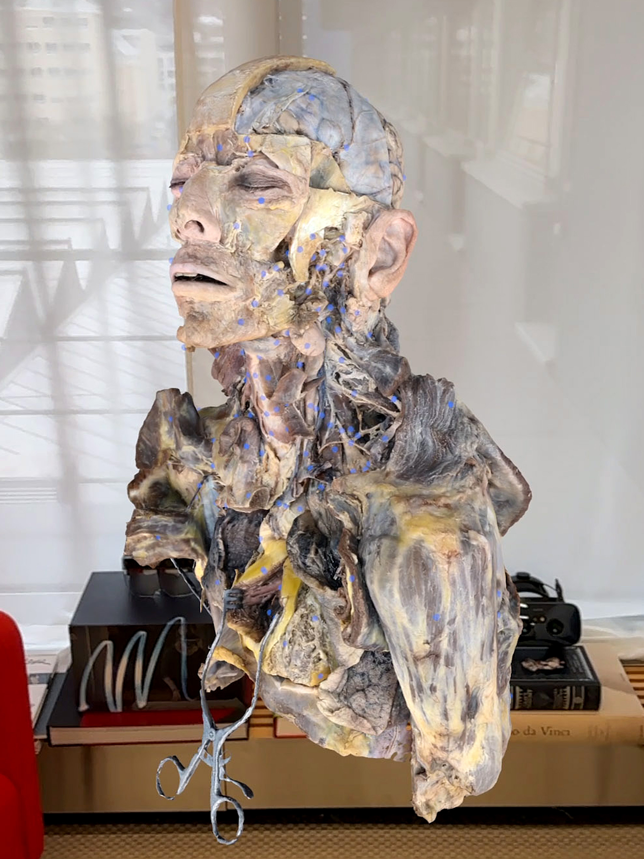
Anatomy Master 2 is now included on standalone devices, PC VR, and Cloud Rendering. Designed for beginners and entry-level learners, these models provide a detailed and realistic view essential for comprehensive understanding.

Anatomy Master 2 offers an interactive and efficient way to learn human anatomy, surpassing traditional screen-based methods and textbooks. Users can walk around, enlarge, dissect, and create cross-sectional views of 3D models for a comprehensive study from any angle.

AI Segmentation
Pancreas & Pancreatic Tumor, Prostate & Spleen
Benefit from the advanced segmentation capabilities of Medicalholodeck AI, offering precise and detailed analysis for various medical imaging modalities. These models enable accurate imaging, supporting users in identifying and analyzing specific anatomical structures and pathological conditions with greater accuracy and efficiency.

Segmentation for MR Images
Available both locally and via the cloud, this feature provides robust segmentation for MR images, improving workflow and accuracy in medical imaging tasks. By leveraging advanced AI algorithms, TotalSegmentator ensures high-quality segmentation, facilitating better diagnosis, treatment planning, and monitoring of patient outcomes.


Visualization Enhancements
Transfer Functions per Segment
Customize each AI-segmented structure with its own transfer function, allowing for tailored visualization and improved clarity in medical imaging. This feature enhances the ability to differentiate between various tissues and structures, providing a more detailed and insightful view that can aid in diagnosis and treatment planning.

UI Improvements
Manage visibility, opacity, and cut-plane settings for datasets, particularly beneficial for PET/CT merges. This enhancement offers greater control and precision in viewing complex datasets, enabling users to isolate and examine specific areas of interest with ease.

Enhanced PET/CT Rendering
Experience improved visual quality and detail in PET/CT merged images, facilitating better analysis and interpretation of medical scans. This improvement ensures that subtle differences and abnormalities are more easily detectable, enhancing the diagnostic capabilities of PET/CT imaging.
New Features

Teleporting
Navigate effortlessly within the virtual space, enhancing the user experience and making it easier to explore and interact with 3D models. This feature allows users to quickly move to different areas within the virtual environment, improving the efficiency and intuitiveness of virtual reality interactions.

Spatial Anchors
Collaborate effectively using shared spatial anchors, ensuring that all users in a session have a consistent view and interaction with virtual objects. This feature for Quest 3 enhances the collaborative experience in educational and professional settings, allowing multiple users to engage with the same virtual content simultaneously.

Spanish Language Version
Access Dissection Master XR and Anatomy Master 2 contents in Spanish, broadening accessibility and usability for Spanish-speaking users.

Save 3D Model State
Save and manage different states of your 3D models, enabling you to quickly return to specific configurations and streamline your workflow. This feature allows users to easily track and revisit specific model setups, facilitating more organized and efficient use of the platform.

Export and Import Color Presets
Share and apply custom settings easily by exporting and importing transfer functions, enhancing collaboration and consistency. This functionality simplifies the process of transferring specific visualization settings between users and projects, ensuring a standardized approach to data interpretation.

File Management and Performance
Volume Data Resolution Control
Adjust volume data resolution to optimize performance on lower-end devices, ensuring smooth and efficient operation across a range of hardware. This feature allows users to balance visual quality and performance based on their device capabilities, providing a more accessible experience.

Peer-to-Peer File Upload
Introduced with an option to enforce this transfer method, ensuring secure and direct file sharing between users. This method enhances data privacy and security, allowing users to transfer files without relying on intermediary servers.

Editable Shapes
Modifying shapes even after creation offers flexibility and control in customizing models and visualizations. This feature allows users to refine and adjust their models as needed, improving the accuracy and utility of their visualizations.

Resizable PDFs
Adjust the size of PDFs within the platform to fit your needs, enhancing readability and usability. This flexibility ensures that users can view and interact with PDF documents more comfortably and efficiently.

GLB/GLTF Uploads
Upload GLB/GLTF files directly via medicalholodeck.com/link, simplifying the process of adding new models and content to the platform. This feature streamlines the workflow for incorporating external 3D models, enhancing the versatility and expandability of the platform.

PACS Improvements

C-Move DICOM Operation Support for PACS
Added support for PACS with C-Move DICOM operations, enhancing the integration and functionality of medical imaging workflows. This support streamlines the transfer and management of DICOM files, improving the efficiency of medical data handling and analysis.

DICOM Tag Arguments for PACS Requests
Customize PACS requests with DICOM tag arguments, providing greater flexibility and control in accessing and managing medical data. This feature allows users to tailor their data retrieval processes to specific needs, improving the precision and relevance of the retrieved information.

Selectable PACS DICOM Download Protocol
Choose the preferred download protocol for PACS DICOM, optimizing the transfer process based on specific needs and requirements. This flexibility ensures that users can select the most efficient and reliable method for their particular workflow and network conditions.
MEDICALHOLODECK®. Surgery, Anatomy, Medicine, Education, and DICOM Imaging in VR. For Medical Students, Nurses, Doctors and Surgeons. - nooon34
Medicalholodeck is launching RecordXR Studio, designed to simplify and enhance AR and VR content editing. RecordXR Studio provides a powerful platform for creating high-quality 4D content, suitable for educators, developers, and VR creators.


Key Editing Features
RecordXR Studio offers tools for editing your immersive narratives. You can cut, shorten, and stitch your recordings, similar to traditional video editing software. The platform also supports seamless audio editing and synchronization, allowing for detailed adjustments to your projects.
One notable feature of RecordXR Studio is the ability to integrate avatars, adding an interactive element to your content. Additionally, you can create multilingual versions by adding audio tracks in different languages, making your content accessible to a broader audience.
Article Header Image

Streamlined Content Creation and Management
With RecordXR Studio, producing AR and VR content is straightforward. Capture your experiences using the RecordXR app, whether you're developing educational materials, simulations, or other immersive experiences. Utilize your data or Medicalholodeck's extensive assets to create compelling content. Import your recordings into RecordXR Studio to refine and perfect them.
The RXR file format simplifies the recording and editing process, ensuring high-quality results for educational libraries, virtual reality simulations, or detailed surgical training. RXR files are compatible with standalone VR devices like Quest 3, 2, Pro, and tethered PC VR systems, ensuring seamless integration across platforms.

Enhancing Immersive Storytelling
RecordXR Studio supports the creation and management of simulations, classes, or unique projects. Combine your expertise with immersive datasets and encapsulate your knowledge in AR and VR formats for easy storage and sharing worldwide.
Article Header Image

Get Started Today
Request a demo to explore how our solutions can improve your AR and VR content creation process.
For more information or support, contact us at support@medicalholodeck.com or call +1 347 480 1356. Stay updated on product developments, regulatory progress, and availability by subscribing to our newsletter.

For more information, contact info@medicalholodeck.com
MEDICALHOLODECK®. Surgery, Anatomy, Medicine, Education, and DICOM Imaging in VR. For Medical Students, Nurses, Doctors and Surgeons. - nooon34

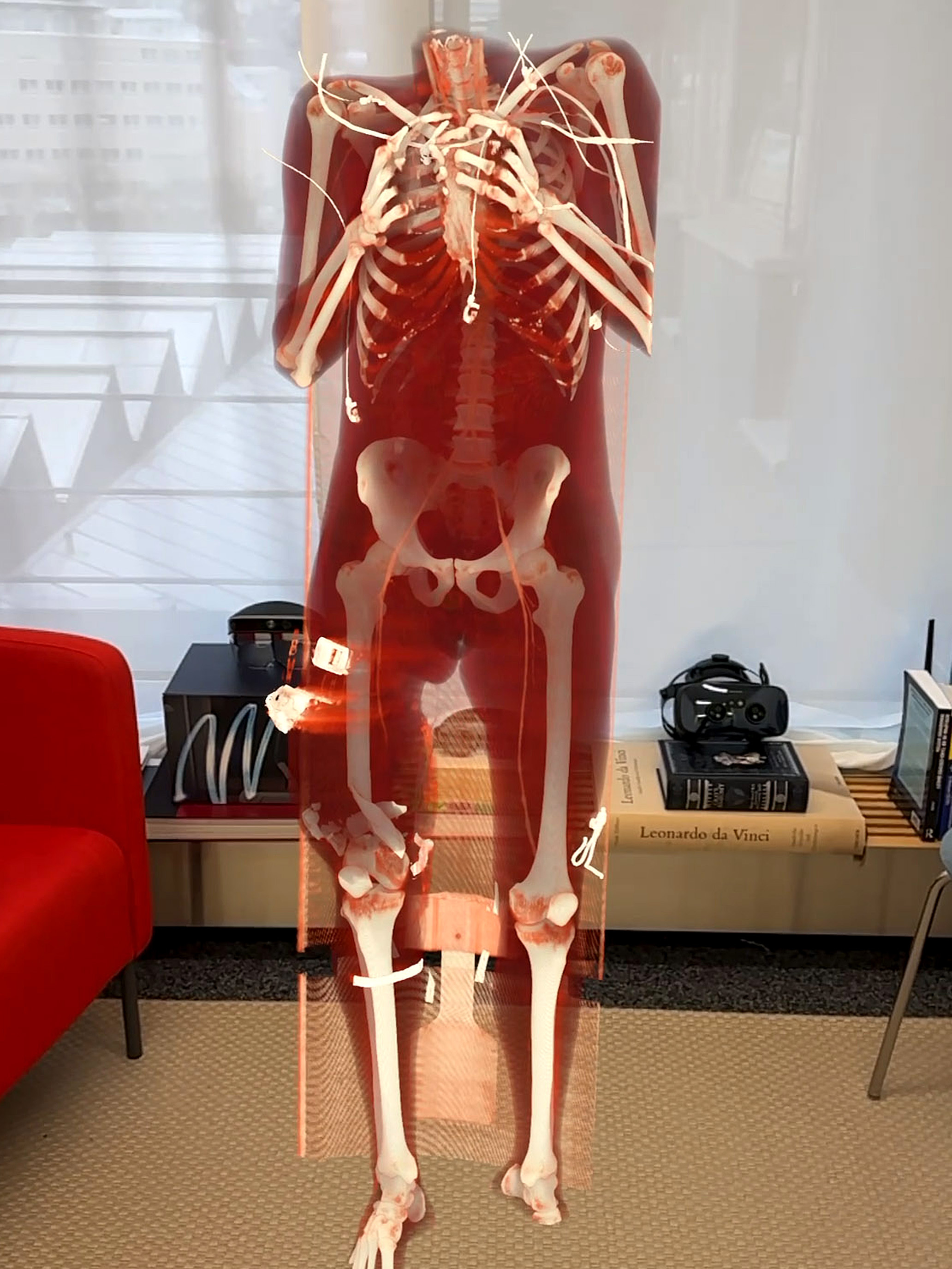
Medicalholodeck is expanding its Medical Imaging software by integrating Positron Emission Tomography (PET) imaging. This update enables seamless integration of PET DICOM files with CT DICOM files, along with AI-powered automatic data segmentation. This enhancement provides a multi-dimensional view of diagnostic data, underlining our commitment to offering the most advanced tools in spatial medical imaging.

Seamless Integration of PET, CT, and AI
PET, a highly valuable imaging technique in various medical specialties, including oncology and neurology, allows clinicians to observe metabolic processes within the body. When fused with CT scans, PET provides not only anatomical details but also metabolic activity, offering a comprehensive overview of a patient's physiological state. Medicalholodeck goes one step further, by making PET, CT, and automatically segmented smart layers easily accessible in AR and VR.

Request a Demo

Automatic Layering, Precise Image Registration
The process starts by importing the PET DICOM files into the Medicalholodeck software, a step as straightforward as uploading any standard dataset. Once uploaded, our innovative layering functionality activates, automatically aligning the PET images with the corresponding CT scans. This ensures precise registration and optimal visualization of the combined datasets. Users can easily switch between PET, CT, or the auto-segmentation views, providing unprecedented insights never before seen.


Advanced Visualization and Easy Control
Medicalholodeck's layering system and automatic alignment algorithm are pivotal for precise analysis and diagnosis. They enable medical professionals to directly correlate metabolic activity with anatomical structures, enhancing both understanding and accuracy. This layering functionality not only streamlines the integration of data but also improves the reliability and utility of the visual output. Users can customize the visualization to meet their specific needs, adjusting layers to achieve the clearest and most informative view possible.

Unparalleled 3D Views for Spatial Medicine
Control over visualization is a cornerstone of the Medicalholodeck experience. Users have the flexibility to adjust each layer individually—fading layers in or out, turning them on or off, and even making precise cuts in one or both layers. Furthermore, individual organs can be isolated, with other structures faded out or hidden, offering unparalleled insights into the depths of your critical medical data. This level of control is crucial in situations where precision and detail are paramount, especially in complex cases where high-quality imaging can directly impact patient outcomes.

3D Imaging for Advanced Healthcare
The ability to manipulate these layers provides an unprecedented 3D view that is not just visually impressive but also immensely practical. Whether for educational purposes, pre-surgical planning, or ongoing patient management, the detailed images rendered in Medicalholodeck facilitate a deeper understanding and a more targeted approach to healthcare.*

*Important Notice Regarding Functionalities for Surgical Planning and Professional Clinical Usage
The specialized features tailored for surgical planning and pre-operative professional applications are exclusive to Medical Imaging XR PRO. At the moment, Medicalholodeck's applications are only available for educational purposes.
MEDICALHOLODECK®. Surgery, Anatomy, Medicine, Education, and DICOM Imaging in VR. For Medical Students, Nurses, Doctors and Surgeons. - nooon34
The integration of virtual reality (VR) and mixed reality (MR) into surgical practices marks a significant advancement, especially in the realm of endoscopic dacryocystorhinostomy (DCR). This article delves into the pioneering work of Rafal Nowak, MD, Izabela Nowak-Gospodarowicz, MD, Marek Rękas, MD, and Mohammad J. Ali, MD, PhD, FRCS, showcasing the transformative potential of these technologies in addressing extremely complex lacrimal drainage obstructions.

https://pubmed.ncbi.nlm.nih.gov/38511475/

The Study: VR and MR in Powered Endoscopic DCR
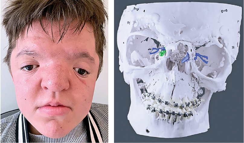
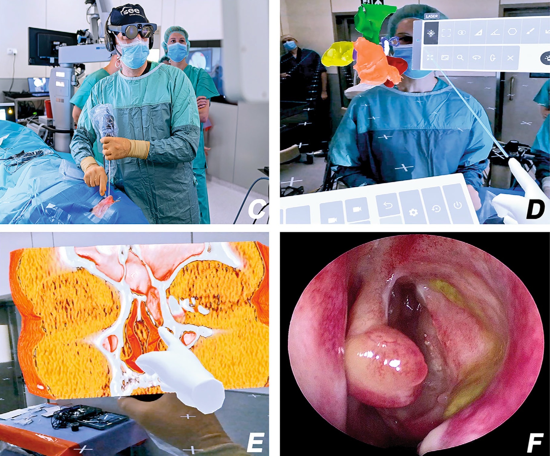
The focal point of this discussion is a study that demonstrates the application of VR and MR in powered endoscopic DCR, particularly targeting complex syndromic congenital nasolacrimal duct obstruction (CNLDO) and post-traumatic secondary acquired lacrimal duct obstruction (SALDO) in patients with Le Fort fractures. This segment explores the methodology and the innovative approach of utilizing preoperative planning with VR and intraoperative guidance with MR to enhance surgical precision.


Case Studies: Overcoming Anatomical Challenges
This section presents detailed case studies from the study, illustrating the effectiveness of VR and MR in navigating the surgical complexities presented by patients with conditions like Apert syndrome and traumatic SALDOs. It highlights how VR models facilitated preoperative assessment and planning, while MR models provided real-time intraoperative guidance, enabling surgeons to navigate through the altered anatomical landscapes with unprecedented precision.
The Impact and Potential of VR and MR in Surgery
The results of this study underscore the viability of VR and MR-assisted powered endoscopic DCR for managing exceptionally challenging cases of CNLDO and SALDO. This part of the article discusses the broader implications of these technologies in surgical practices, emphasizing the enhanced level of anatomical insight and surgical precision they offer.


Challenges and Future Directions
Despite the promising outcomes, the integration of VR and MR into surgical procedures comes with its set of challenges, including additional costs, the need for extensive planning, a steep learning curve, and the requirement for specialized equipment. This section addresses these challenges and considers the future of VR and MR in surgical practices, highlighting the need for further research and clinical trials to fully harness the potential of these innovative technologies.

Conclusion: A New Horizon in Surgical Practice
The article concludes by reaffirming the significant contributions of Nowak, Nowak-Gospodarowicz, Rękas, and Ali in integrating VR and MR into endoscopic DCR procedures. It reflects on the transformative impact of these technologies on complex lacrimal surgeries and anticipates their broader application in advancing surgical practices across various disciplines.

https://www.medicalholodeck.com/en/
...

Search news
Archive
2025
May   Apr   Mar   Feb   Jan  
Archives By Year
2025   2024   2023   2022   2021  
2020   2019   2018   2017   2016  
2015   2014   2013   2012   2011  
2010   2009   2008   2007   2006  
2005   2004   2003   2002