Mar 8, 2024
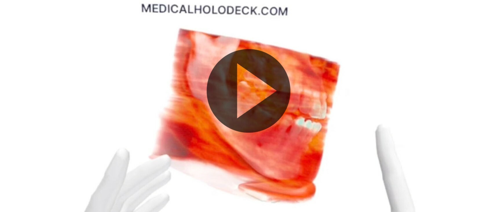
MEDICALHOLODECK®. Surgery, Anatomy, Medicine, Education, and DICOM Imaging in VR. For Medical Students, Nurses, Doctors and Surgeons. - nooon34
Learn from leading experts how Medicalholodeck improves medicine and education. Understand how AI is transforming medical images into smart data, and learn about the impact in pediatrics and neurosurgery. Join us on March 22!
Website + Registration: https://www.medicalholodeck.com/demo-day-2024

We will cover the latest in Medicalholodeck technology, showcasing AI's role in transforming it into a smart platform for professionals and how RecordXR is revolutionizing medical education and content creation. Experts from Europe and the U.S. will share their experiences with Medicalholodeck's impact on incorporating virtual reality in medicine.
CET: 12.45 | EDT: 7.45am | CDT: 6.45am | PDT: 4.45am
What’s Next on Deck? Welcome Address by the Founders
Presenting on stage at Technopark Zurich, Switzerland + OnlineChristof (CEO), Dominique (CTO)
CET: 13.00 | EDT: 8am | CDT: 7am | PDT: 5am
3D Reconstruction Techniques To Augment the Planning of Complex Pediatric Surgical Procedures
Online from Dallas, Texas.Mark Ryan, MD, MSPH, FACS | Assistant Professor | Surgical Medical Director, ECMO | Surgical Medical Director, Trauma ICU | Associate Medical Director, Trauma Services | Department of Surgery, Division of Pediatric Surgery | UT Southwestern Medical Center/Children's Medical Center Dallas
CET: 13.30 | EDT: 8.30am | CDT: 7.30am | PDT: 5.30am
Medical XR in Thoracic Surgery. AI and Spatial Imaging Infrastructure in a Leading German Hospital
Presenting on stage at Technopark Zurich, Switzerland + OnlineJan C. Arensmeyer M.D., and Philipp Feodorovici M.D., Bonn Surgical Technology Center, University Hospital Bonn, Germany
CET: 14.00 | EDT: 9am | CDT: 8am | PDT: 6am
Spatial Computing in Spine Surgery: Past, Present, Future
Online from New York City.Galal A. Elsayed M.D. Assistant Professor and Neurosurgeon, Weill Cornell Medicine at New York-Presbyterian, Queens-Och Spine
CET: 14.30 | EST: 8.30am | CT: 7.30am | PST: 5.30am
Creating the World's Most Stunning XR Anatomy Experience: Meet the Dissection Master
Presenting on stage at Technopark Zurich, Switzerland + OnlineEvan Goldman, Ph.D., Associate Professor | Director of Anatomy | Director of the Willed Body Program | Department of Radiology | Penn State College of Medicine, Hershey, PA
CET: 15.00 | EST: 9am | CT: 8am | PST: 6am
Virtual Reality Unmasked: Reconstructing a Cranial Axe Attack from a Neurosurgical Perspective
Presenting on stage at Technopark Zurich, Switzerland + OnlineMarkus Holling, MD, PhD, MHBA | Professor & Deputy Chairman | Department of Neurosurgery | University Hospital Münster, Germany
Website + Registration: https://www.medicalholodeck.com/demo-day-2024
Share: